An In-Depth Look at Electrocardiography (ECG)
The Basics of Electrocardiography (ECG): Electrocardiography, commonly referred to as ECG or EKG (from the German “Elektrokardiogramm”), is a non-invasive medical technique that records the electrical activity of the heart over time. The heart’s contractions are driven by electrical signals generated by specialized cells within its walls. By placing electrodes on the skin’s surface, ECG captures these electrical impulses and translates them into visual representations known as electrocardiograms.
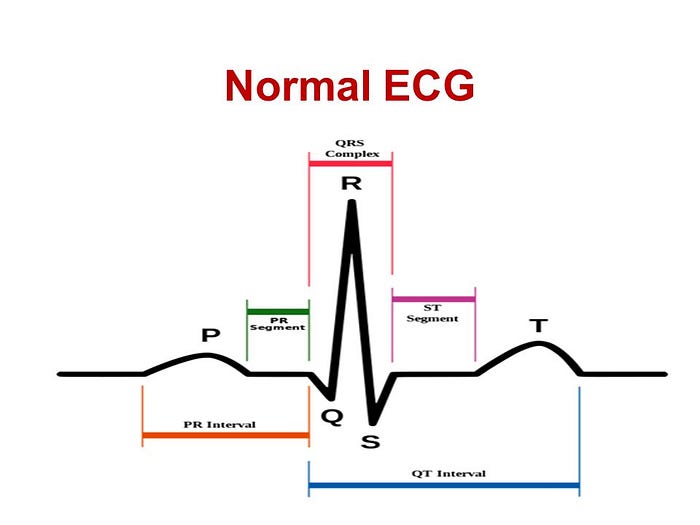
An ECG tracing consists of several distinct waves and intervals, each corresponding to a specific event in the cardiac cycle. The P wave signifies atrial depolarization, the QRS complex represents ventricular depolarization, and the T wave denotes ventricular repolarization.

Einthoven Triangle
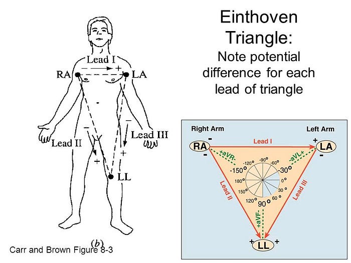
The Einthoven Triangle is a fundamental concept in electrocardiography (ECG). It is named after Willem Einthoven, a Dutch physiologist who contributed significantly to the development of ECG technology. The Einthoven Triangle is not a physical triangle; rather, it is a theoretical construct that represents the arrangement of the limb electrodes used in a standard ECG.
The Einthoven Triangle involves three limb electrodes placed on the body:
- Lead I (RA — LA): This lead records the voltage between the right arm (RA) and the left arm (LA).
- Lead II (RA — LL): This lead records the voltage between the right arm (RA) and the left leg (LL).
- Lead III (LA — LL): This lead records the voltage between the left arm (LA) and the left leg (LL).

These three leads form the vertices of the Einthoven Triangle, even though they don’t form a physical triangle on the body. By measuring the electrical potential differences between these different limb electrodes, medical professionals can obtain crucial information about the heart’s electrical activity and diagnose various cardiac conditions.
The Einthoven Triangle is a cornerstone of ECG interpretation, and it helps in understanding the electrical axis of the heart, as well as detecting and diagnosing arrhythmias, ischemia, and other heart-related issues.
Types of ECG:
- Resting ECG (Standard ECG): This is the most common form of ECG, typically performed while the patient is at rest. Electrodes are placed on specific locations on the chest, arms, and legs to record the heart’s electrical activity over a short period. Resting ECG is crucial for diagnosing conditions like arrhythmias, ischemia, and structural abnormalities.
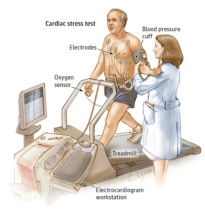
- Stress ECG (Exercise ECG or Treadmill Test): In this type, the patient’s heart activity is monitored while they engage in controlled physical exercise, usually on a treadmill or stationary bicycle. Stress ECG is used to assess the heart’s response to exertion, helping diagnose coronary artery disease and evaluating exercise capacity.

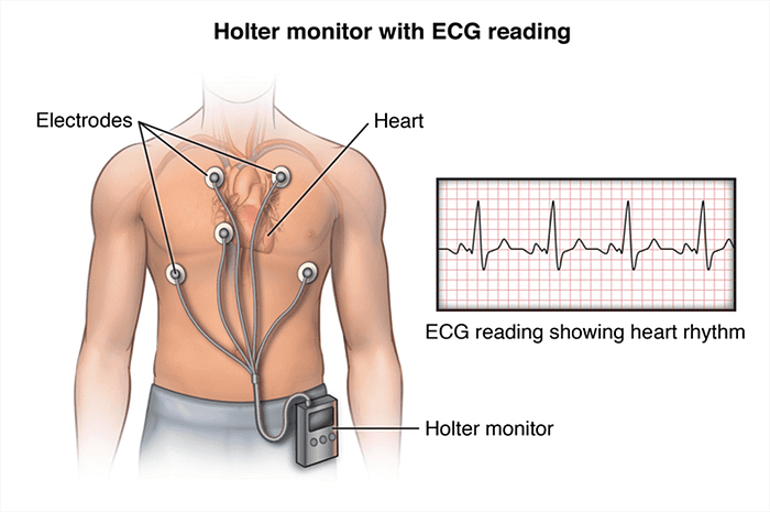
Holter Monitoring: This involves continuous ECG recording over a prolonged period, often 24 to 48 hours, while the patient goes about their daily activities. Holter monitoring is invaluable for capturing intermittent arrhythmias and identifying potential triggers.

Event Monitor: Similar to a Holter monitor, an event monitor is worn by the patient for longer durations. However, it is activated by the patient when they experience symptoms, allowing for targeted monitoring during specific events.
Ambulatory ECG (Ambulatory Holter Monitoring): This type involves prolonged ECG recording, often for several days, using a small wearable device. Ambulatory ECG provides a comprehensive assessment of the heart’s activity during various activities and sleep.
Signal-Averaged ECG: This technique involves averaging multiple ECG complexes to enhance the detection of subtle abnormalities, particularly useful for identifying ventricular arrhythmias.
Vectorcardiography: Vectorcardiograms provide a spatial representation of the heart’s electrical activity by measuring the magnitude and direction of electrical vectors. This advanced technique offers insights into complex arrhythmias and conduction abnormalities.
Phonocardiography (PCG): Phonocardiography is the recording and analysis of sounds produced by the heart, particularly the heart sounds
Electrocardiography is a valuable diagnostic tool used to detect and diagnose a variety of heart-related conditions and abnormalities. Here are some of the diseases and conditions that can be diagnosed or indicated through ECG:
- Arrhythmias: ECG is particularly effective in diagnosing arrhythmias, which are irregular heart rhythms.
- Myocardial Infarction (Heart Attack): ECG can reveal signs of reduced blood flow to the heart muscle, indicating a heart attack. Changes in the ST segment and the presence of Q waves are characteristic findings in acute myocardial infarction.
- Ischemia: ECG changes can indicate myocardial ischemia, which is reduced blood flow to the heart muscle. This can be a precursor to a heart attack. Changes in the ST segment during exercise (stress test) or under resting conditions may suggest ischemia.
- Cardiomyopathies: ECG can provide insights into various types of cardiomyopathies, such as hypertrophic cardiomyopathy, dilated cardiomyopathy, and restrictive cardiomyopathy.
- Pericarditis: Inflammation of the pericardium, the sac surrounding the heart, can cause characteristic changes on the ECG, such as diffuse ST segment elevation and PR segment depression.
